
AlphaFold predicted structure
AP2M1 · Q96CW1
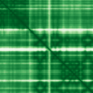
Mean pLDDT
89.2/ 100
Confident
435 residues
Confidence breakdown
- Very high(≥ 90)72%
- Confident(70–90)21%
- Low(50–70)4%
- Very low(< 50)3%
Open interactive 3D viewer
AlphaFold (Jumper et al., 2021) · CC BY 4.0
adaptor related protein complex 2 subunit mu 1
Annotations refreshed 2 months ago.
Diagnostic Grade (Green)
DDG2P
MONOALLELIC, autosomal or pseudoautosomal, imprinted status unknownEarly onset or syndromic epilepsy
MONOALLELIC, autosomal or pseudoautosomal, imprinted status unknownIntellectual disability
MONOALLELIC, autosomal or pseudoautosomal, imprinted status unknownintellectual developmental disorder 60 with seizures
neurodegenerative disease
HIV infectious disease
lysosomal storage disease
Alzheimer disease
Parkinson disease
Intellectual disability
Global developmental delay
developmental and epileptic encephalopathy
Ataxia
Score is the Open Targets composite evidence score (0-1). Higher = stronger gene-disease association.
AP-2 complex subunit mu
Component of the adaptor protein complex 2 (AP-2) (PubMed:12694563, PubMed:12952941, PubMed:14745134, PubMed:14985334, PubMed:15473838, PubMed:31104773). Adaptor protein complexes function in protein transport via transport vesicles in different membrane traffic pathways (PubMed:12694563, PubMed:12952941, PubMed:14745134, PubMed:14985334, PubMed:15473838, PubMed:31104773). Adaptor protein complexes are vesicle coat components and appear to be involved in cargo selection and vesicle formation (PubMed:12694563, PubMed:12952941, PubMed:14745134, PubMed:14985334, PubMed:15473838, PubMed:31104773). AP-2 is involved in clathrin-dependent endocytosis in which cargo proteins are incorporated into vesicles surrounded by clathrin (clathrin-coated vesicles, CCVs) which are destined for fusion with the early endosome (PubMed:12694563, PubMed:12952941, PubMed:14745134, PubMed:14985334, PubMed:15473838, PubMed:31104773). The clathrin lattice serves as a mechanical scaffold but is itself unable to bind directly to membrane components (PubMed:12694563, PubMed:12952941, PubMed:14745134, PubMed:14985334, PubMed:15473838, PubMed:31104773). Clathrin-associated adaptor protein (AP) complexes which can bind directly to both the clathrin lattice and to the lipid and protein components of membranes are considered to be the major clathrin adaptors contributing the CCV formation (PubMed:12694563, PubMed:12952941, PubMed:14745134, PubMed:14985334, PubMed:15473838, PubMed:31104773). AP-2 also serves as a cargo receptor to selectively sort the membrane proteins involved in receptor-mediated endocytosis (PubMed:16581796). AP-2 seems to play a role in the recycling of synaptic vesicle membranes from the presynaptic surface (PubMed:12694563, PubMed:12952941, PubMed:14745134, PubMed:14985334, PubMed:15473838, PubMed:31104773). AP-2 recognizes Y-X-X-[FILMV] (Y-X-X-Phi) and [ED]-X-X-X-L-[LI] endocytosis signal motifs within the cytosolic tails of transmembrane cargo molecules (By similarity). AP-2 may also play a role in maintaining normal post-endocytic trafficking through the ARF6-regulated, non-clathrin pathway (PubMed:19033387). During long-term potentiation in hippocampal neurons, AP-2 is responsible for the endocytosis of ADAM10 (PubMed:23676497). The AP-2 mu subunit binds to transmembrane cargo proteins; it recognizes the Y-X-X-Phi motifs (By similarity). The surface region interacting with to the Y-X-X-Phi motif is inaccessible in cytosolic AP-2, but becomes accessible through a conformational change following phosphorylation of AP-2 mu subunit at Thr-156 in membrane-associated AP-2 (PubMed:11877457). The membrane-specific phosphorylation event appears to involve assembled clathrin which activates the AP-2 mu kinase AAK1 (PubMed:11877457). Plays a role in endocytosis of frizzled family members upon Wnt signaling (By similarity)
Curated MONDO disease pages that list AP2M1 among their top associated genes.
AP2M1 · Q96CW1
Mean pLDDT
89.2/ 100
Confident
435 residues
Confidence breakdown
AlphaFold (Jumper et al., 2021) · CC BY 4.0