Skip to content
GenoLensGenoLens

IRAK4

Chr 12q12

interleukin 1 receptor associated kinase 4

Aliases:
NY-REN-64
MANE:
ENST00000613694.5

Annotations refreshed 9 hours ago.

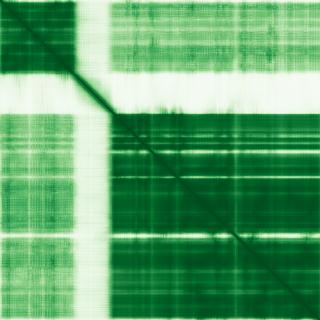
Predicted protein structure

Clinical relevance (Genomics England PanelApp)

Diagnostic Grade (Green)

  • COVID-19 research

    BIALLELIC, autosomal or pseudoautosomal
  • Primary immunodeficiency or monogenic inflammatory bowel disease

    BIALLELIC, autosomal or pseudoautosomal

Disease associations (Open Targets)

  • Immunodeficiency due to interleukin-1 receptor-associated kinase-4 deficiency

    0.73
  • immunodeficiency 67

    0.73
  • inborn error of immunity

    0.46
  • autoimmune disorder of central nervous system

    0.37
  • immunodeficiency 33

    0.33
  • congenital dyserythropoietic anemia

    0.30
  • pericarditis

    0.25
  • neurodegenerative disease

    0.25
  • hereditary disease

    0.19
  • alcohol drinking

    0.12

Score is the Open Targets composite evidence score (0-1). Higher = stronger gene-disease association.

Protein function (UniProt)

Interleukin-1 receptor-associated kinase 4

Serine/threonine-protein kinase that plays a critical role in initiating innate immune response against foreign pathogens. Involved in Toll-like receptor (TLR) and IL-1R signaling pathways (PubMed:17878374). Is rapidly recruited by MYD88 to the receptor-signaling complex upon TLR activation to form the Myddosome together with IRAK2. Phosphorylates initially IRAK1, thus stimulating the kinase activity and intensive autophosphorylation of IRAK1. Phosphorylates E3 ubiquitin ligases Pellino proteins (PELI1, PELI2 and PELI3) to promote pellino-mediated polyubiquitination of IRAK1. Then, the ubiquitin-binding domain of IKBKG/NEMO binds to polyubiquitinated IRAK1 bringing together the IRAK1-MAP3K7/TAK1-TRAF6 complex and the NEMO-IKKA-IKKB complex. In turn, MAP3K7/TAK1 activates IKKs (CHUK/IKKA and IKBKB/IKKB) leading to NF-kappa-B nuclear translocation and activation. Alternatively, phosphorylates TIRAP to promote its ubiquitination and subsequent degradation. Phosphorylates NCF1 and regulates NADPH oxidase activation after LPS stimulation suggesting a similar mechanism during microbial infections

Data sources: HGNC (CC BY 4.0), AlphaFold (CC BY 4.0, Jumper et al. Nature 2021), Genomics England PanelApp (CC BY 4.0), ClinGen, Open Targets (CC0), UniProt.

Not for sole clinical decision-making. Always verify against primary sources.